Beauty Under the Microscope
Thayer engineering students (mostly Ph.D. candidates) recently produced images to highlight the creativity and beauty of research as part of a Visionaries in Technology contest. Here are a few examples of their extreme close-ups.
Thayer engineering students (mostly Ph.D. candidates) recently produced images to highlight the creativity and beauty of research as part of a Visionaries in Technology contest. Here are a few examples of their extreme close-ups.
Title
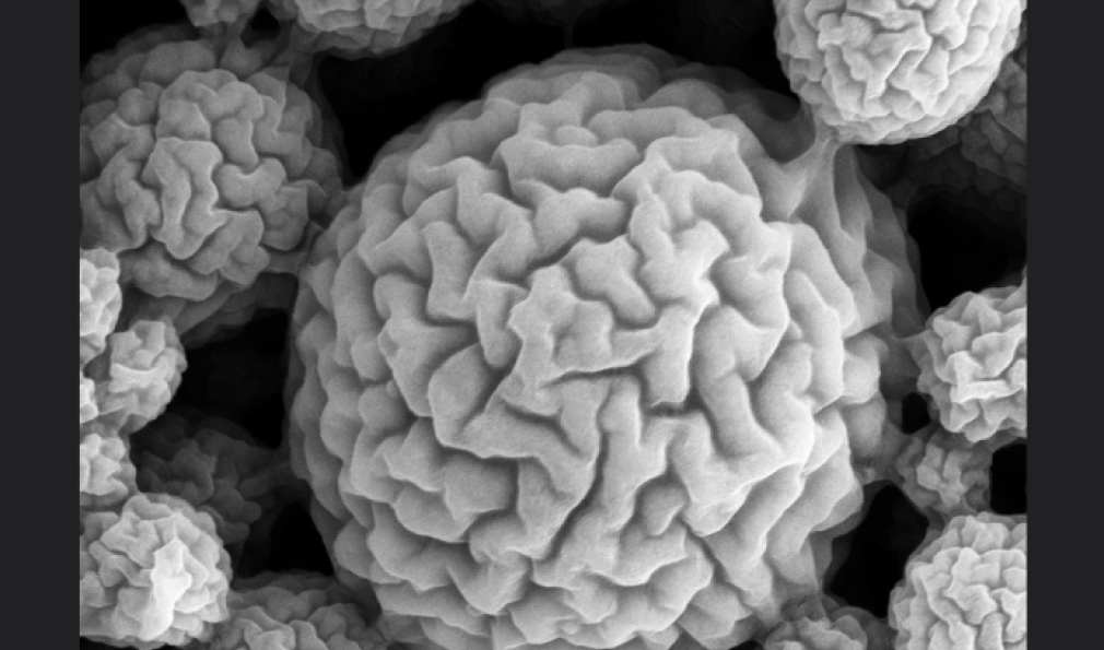
Brainballs
Description
De-hydrated alginate spheres, imaged with the secondary electron detector in a scanning electron microscope (SEM).
Photo Credit
Philipp Hunger
Title
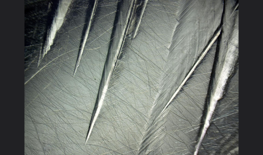
Damage Features on a Metal Hip Implant
Description
Imaged at 100x using a Keyence microscope.
Photo Credit
Steven Reinitz
Title
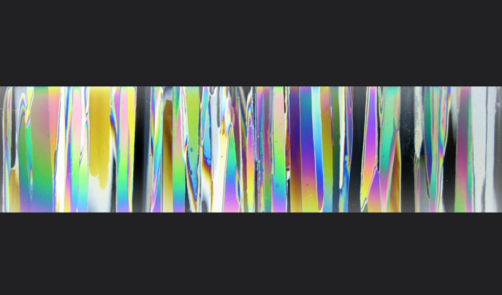
Freshwater Ice Under Microscope
Description
Digital photograph of thin section of polycrystalline S2 freshwater ice under cross-polarized light to illuminate its columnar-grained microstructure.
Photo Credit
Scott Snyder
Title
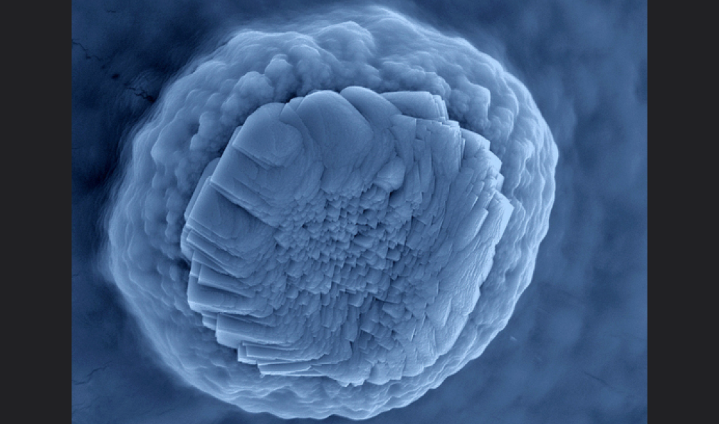
Jelly Fish
Description
Calcium carbonate crystal, false colored with a blue filter.
Photo Credit
Philipp Hunger
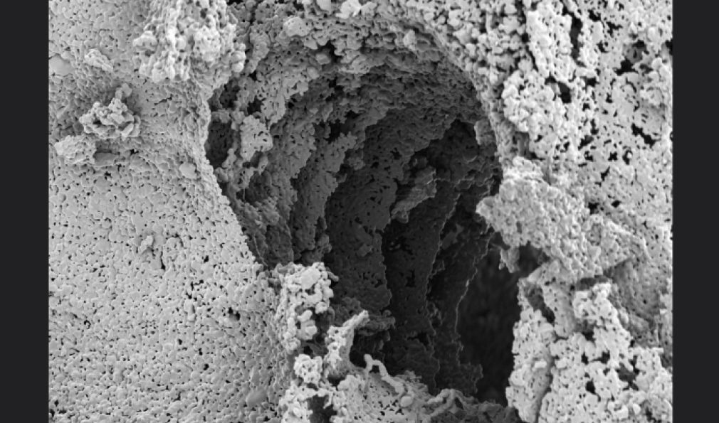
Title
Layer by Layer
Description
Sintered, freeze-cast hydroxyapatite scaffold with horizontal canal, templated by an alginate strut.
Photo Credit
Philipp Hunger
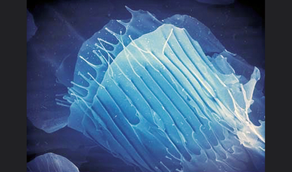
Title
Microridged Scaffolds
Description
Highly aligned microridges run alongside the walls in ice-templated carboxymethylcellulose scaffolds.
Photo Credit
Margaret Wu
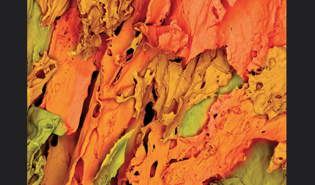
Title
Nano Autumn
Description
Chitosan scaffold imaged by SEM at 100X. First-place winner (tied).
Photo Credit
Fioleda Prifti
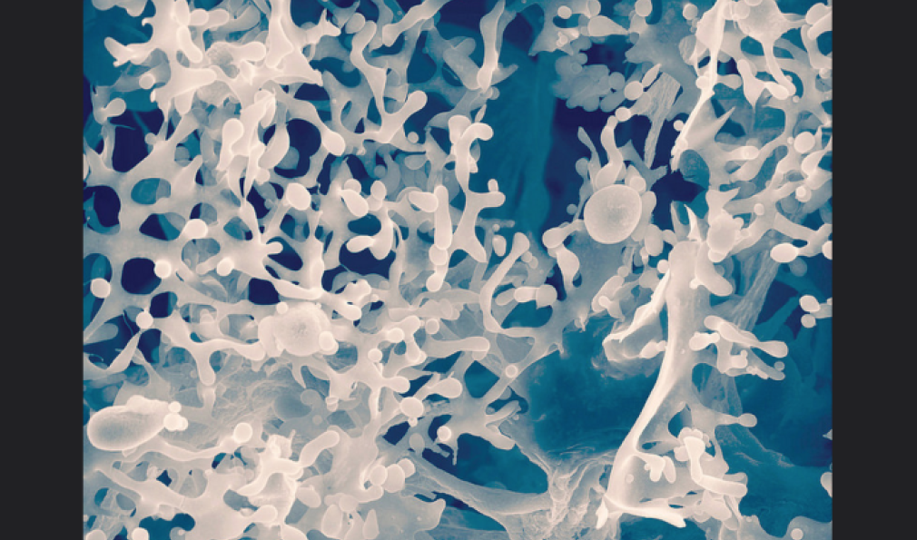
Title
Nano Bloom
Description
Nanocellulose fibril scaffold. Second-place winner (tied).
Photo Credit
Fioleda Prifti
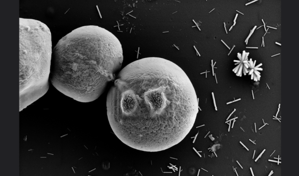
Title
Pacman
Description
Chitosan Spheres with additional “burnmarks” created with the electron beam.
Photo Credit
Philipp Hunger
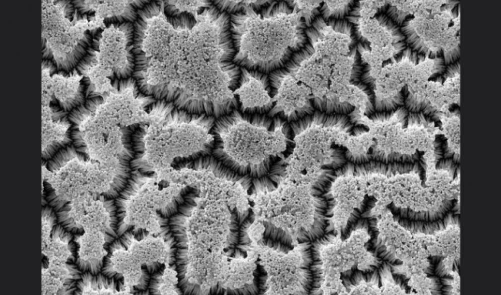
Title
Polyvinylidene Fluoride (PVDF) Vertical Nanofiber Array
Description
PVDF fiber with 300nm diameter and 3um length fabricated by template assisted method. First-place winner (tied).
Photo Credit
Dajing Chen
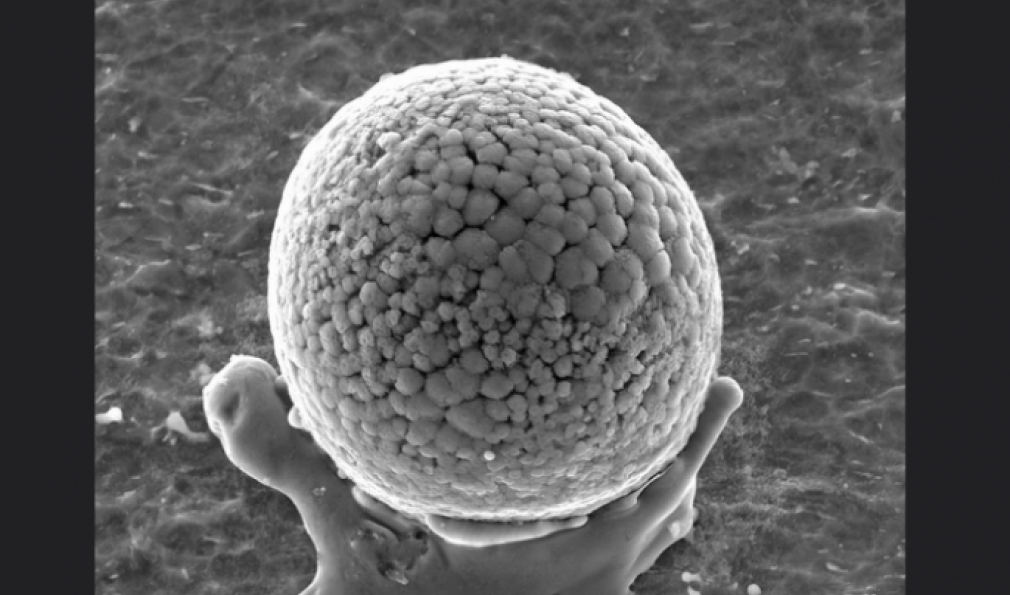
Title
Atlas’ Hand
Description
Debris adhered to the inner surface of a titanium tube was deposited during cutting of the tube and caused detrimental scratches.
Photo Credit
Jay Vincelli


